Techniques
From single molecule to whole muscle#
Myolab uses a multi-scale biophysical approach to study skeletal and cardiac muscle physiology in health and disease. The following are the experimental techniques we use to understand various aspects of muscle contraction, arranged from smallest -> largest biological level.
Single-molecule optical trapping#
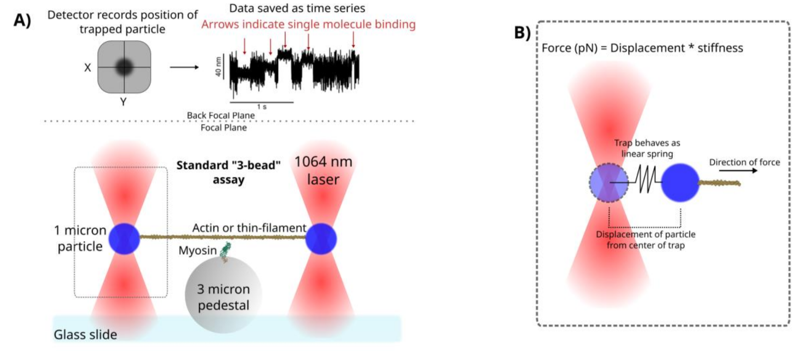
We use single-molecule optical trapping with the “3-bead” geometry to measure the forces generated by individual proteins of the muscle in an isolated and highly controlled in vitro system. For example, with this technique, we can measure the distance a single myosin molecule moves an actin filament each ATPase cycle, which is about 5 nanometers and generates ~1 piconewton of force. We have an Impetux SENSOCELL optical trapping system.
Below is a cartoon representation of the optical trapping experiment which is performed on a customized Nikon TiU inverted microscope. A 1064 nm laser is directed through an optical path and into the microscope. The microscope’s objective focuses the laser light near the focal plane creating an “optical trap” which allows us to grab and manipulate small particles (similar to a tractor beam in SCI-FI movies). We trap 1 micron microspheres which we make “sticky” to actin via a streptavidin-biotin linkage. We adhere an actin filament to the the two trapped microspheres and pull it taught by steering the laser beams. We can then lower the actin filament towards the surface of the microscope slide surface to interact with a single molecule (myosin is depicted here). We have a special detector mounted in the back focal plane of the microscope that records the position of the trapped microspheres. The detector measures both distances and forces with nanometer and piconewton resolution.
Mutli-molecule optical trapping#
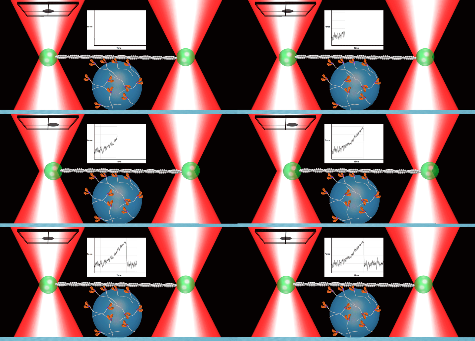
While single molecule experiments provide the fundamental and unitary measurements of single protein interactions, adding additional proteins allows us to begin to recapitulate higher order physiological structures to understand how proteins cooperatively work together to generate force within the muscle. Below is an animation made by the Muscle Biophysics Lab at the University of Massachusetts Amherst to demonstrate a small team of myosin molecules working to displace an actin filaments in the “mini-ensemble” optical trapping experiment.
In vitro motility#
Also known as a “gliding assay” in other labs, we prefer to call this experiment “muscle contraction in a dish” or “crowd-surfing actin”. We stick a bunch of myosin motors to a microscope slide and watch them translocate fluorescently labeled actin filaments over this dense lawn of myosin using epi-fluorescence. This experiment is done a Nikon TiU inverted microscope.
Ex vivo muscle mechanics#
We have an Aurora Scientific 805A isolated muscle bath to perform single muscle mechanics using rodent skeletal muscles (tendon to tendon) or cardiac tissue (papillary or trabeculae).
In vivo muscle mechanics#
We also have an Aurora Scientific 806D which allows us to measure dorsi- and plantar-flexion forces in living rats.